What is Knee Arthroscopy?
Knee arthroscopy is a keyhole procedure that lets an orthopaedic surgeon see inside the knee through small incisions. A camera called an arthroscope sends magnified images to a monitor, while fine instruments can treat selected problems. Knee arthroscopy may be used for certain meniscus tears, loose fragments, ligament reconstruction and cartilage injuries. It is often day surgery, but recovery depends on what is actually treated.
What Is Knee Arthroscopy?
During knee arthroscopy, the joint is gently filled with sterile fluid so the surgeon can inspect structures such as the menisci, articular cartilage and ligaments. The camera provides a clear, enlarged view without the exposure required for traditional open surgery.
Modern imaging has reduced the need for knee arthroscopy purely for diagnosis. Examination, X-rays and MRI often provide substantial information first. Arthroscopy is most useful when symptoms and imaging point to a problem that can genuinely be assessed or treated through the scope.
What Can Knee Arthroscopy Treat?
Knee arthroscopy surgery may be considered for:
- repairing or trimming selected meniscus tears
- removing loose pieces of cartilage or bone
- treating selected cartilage lesions
- reconstructing a torn ligament, including the ACL
- removing problematic scar or inflamed tissue in selected cases
- assessing and treating certain problems that remain unresolved after clinical examination and imaging.
Not every painful knee needs an arthroscope. In particular, arthroscopy has a limited role for pain caused mainly by established knee osteoarthritis. Treatment should be matched to the actual cause of symptoms rather than the presence of pain alone.
How Is a Knee Arthroscopy Procedure Performed?
Small portals are made around the knee. The arthroscope, which contains a lens and light system, is introduced through one portal. Images are displayed on a monitor, allowing the surgeon to inspect the joint systematically.
If treatment is needed, small instruments pass through additional portals. A shaver can remove selected damaged tissue, while repair or reconstruction instruments are used when indicated. The steps vary considerably between a simple procedure, meniscus repair and ACL reconstruction.
What Anaesthesia Is Used?
Knee arthroscopy can be performed under general, spinal or, in selected situations, local anaesthesia. The choice depends on the planned procedure, general health and the anaesthetist’s assessment.
NHS guidance notes that arthroscopy usually takes between 30 minutes and two hours, depending on what is being done. Many patients go home after recovering from the anaesthetic, although an overnight stay can occasionally be necessary.
Who May Not Be Suitable for Knee Arthroscopy?
Knee arthroscopy is not automatically appropriate because an MRI shows a tear or degeneration. Surgery may be postponed when there is active infection, significant uncontrolled illness or a skin problem near the operative area. It may also offer little value when symptoms are primarily caused by advanced osteoarthritis and no treatable mechanical problem has been identified.
For some meniscus injuries, physiotherapy and activity modification may be reasonable first options. The decision depends on the tear pattern, symptoms, age, activity demands, associated injuries and whether the knee locks or remains unstable.
Knee Arthroscopy Recovery Time
Knee arthroscopy recovery time varies widely because “arthroscopy” describes an approach, not one operation. NHS guidance gives a broad overall range from about one week to several months. A minor procedure may settle relatively quickly, while meniscus repair, cartilage work or ligament reconstruction usually requires a more structured rehabilitation period.
Pain, swelling and stiffness are expected early. Crutches may be needed briefly or for longer when weight-bearing is restricted. General health, muscle strength, the tissue repaired and rehabilitation all affect recovery.
Walking, Stairs, Driving and Work
Walking often begins on the day of surgery, with crutches when required. Instructions about weight-bearing should come from the surgical team because they change according to the procedure performed.
Driving should wait until the patient can control the vehicle safely and perform an emergency stop, and is no longer affected by anaesthetic or medicines that impair alertness. Return to work also varies. Desk work may be possible considerably earlier than a job involving prolonged standing, lifting, climbing or manual labour.
Will Physiotherapy Be Needed?
Rehabilitation is tailored to the procedure. Simple cases may need home exercises, while meniscal repair or ACL reconstruction commonly requires supervised rehabilitation. Early priorities include swelling control, movement and quadriceps activation before strength, balance and sport-specific work progress.
What Are the Risks of Knee Arthroscopy?
Knee arthroscopy uses small incisions, but it is still surgery. Recognised complications include infection, bleeding inside the joint, blood clots, anaesthetic reactions and damage to structures within or around the knee. Persistent pain, swelling or stiffness can also occur, particularly when the underlying knee problem cannot be fully corrected arthroscopically.
Seek prompt medical advice for fever, worsening redness or swelling, unusual or unpleasant wound drainage, severe increasing pain, new numbness or tingling, calf swelling, chest pain or breathlessness.
What Are the Alternatives to Knee Arthroscopy?
Alternatives depend on the diagnosis. They may include activity modification, physiotherapy, weight management, prescribed pain relief, injections or observation. Some ligament injuries can initially be managed with rehabilitation, while particular meniscus tears may heal or become manageable without surgery.
For advanced arthritis, joint-preserving procedures, partial knee replacement or total knee replacement may be more appropriate than arthroscopy in selected patients. The important question is not whether knee arthroscopy is minimally invasive, but whether it addresses the source of the symptoms.
What Current Evidence Says About Meniscus Treatment
The approach to meniscus injury has become more tissue-preserving. Current orthopaedic guidance emphasises that treatment depends on tear type, location, symptoms and tissue quality. When a tear is suitable for repair, preserving functioning meniscal tissue may be preferable to removing it unnecessarily.
This is also why a knee arthroscopy procedure should not be described as routine “clean-up”. The surgeon’s aim and the biological problem being treated matter. A repair intended to preserve the meniscus has a different recovery plan from a partial meniscectomy, where damaged tissue is trimmed.
Dr Amyn Rajani’s Research in Knee Arthroscopy
Dr Amyn Rajani’s scientific publications include work directly related to arthroscopic meniscus repair and ACL surgery. His research therefore adds procedure-specific context rather than simply a general reference to his Google Scholar profile.
The AMR Sign and Medial Meniscus Repair
In a 2023 multicentre prospective study, Dr Rajani and colleagues evaluated 804 patients undergoing arthroscopic all-inside repair for isolated medial meniscus tears. The researchers described the AMR, or Adequacy of Medial Meniscus Repair, sign: an S-shaped fold visible at the free inner edge of the repaired meniscus.
Patients in whom the sign formed and remained had better functional outcomes and lower failure rates at terminal follow-up. The study is useful because it evaluated an objective arthroscopic sign of repair adequacy. Its findings apply to the technique and patient group studied, not every knee arthroscopy.
ACL Research and Arthroscopic Reconstruction
Dr Rajani has also published research on ACL reconstruction. A 2022 prospective study followed 113 patients who underwent ACL reconstruction using a peroneus longus autograft and assessed clinical, functional and MRI outcomes after three years. His 2025 research also examined femoral intercondylar notch shape and non-contact ACL tear risk in 100 patients undergoing knee MRI.
Together, these studies show why tissue preservation, anatomy, graft choice and rehabilitation matter alongside the arthroscopic technique itself.
The image by an Arthroscope is magnified and allows the surgeon to see better and clearer. The Arthroscope allows the Arthroscopic Surgeon to see and perform Surgery using much smaller incisions. This results in less tissue damage to normal tissue and can shorten the healing process.
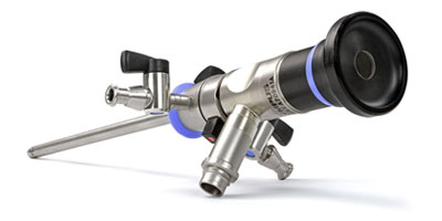
Arthroscope:
An Arthroscope is a cylinder with a lens at each end. A light source is attached to the Arthroscope by a Fiber Optic Cable. The light is shone down the Arthroscope in the same direction as the surgeon is viewing in order to illuminate the interior of the joint.
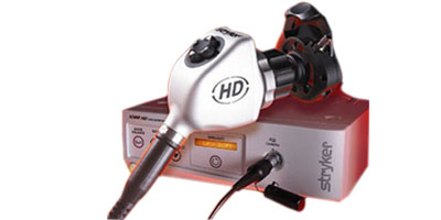
Camera:
The Arthroscope is locked in the camera which is attached to the console. The buttons on the camera can adjust the amount of light. The console in turn is connected to a TV and recorder. The TV will display everything visualized in the joint, and the recorder will record these videos in DVD format.
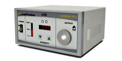
Light Source:
The Fiber Optic Cable connects the Arthroscope to the light source for illumination in the joint. these cables are Water tight for easy cleaning, and soaking in sterilizing solutions.
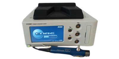
Arthroscopy Shaver:
This is a power device which helps to shave the meniscus, cartilage flaps and fat pad.
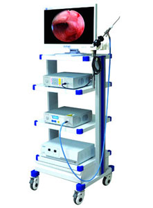
Arthroscopy Console Trolley:
The TV, recorder, camera console, light source and shaver console are placed on a trolley close to the operating table.
Arthroscopy is a day care procedure where in the patient is admitted in the hospital on same day in the morning and is discharged on the same day in the evening. As there is very little interference with body systems as well as the joint tissues the patient not only goes home the same day but is also able to start regaining body functions by same evening or max by next day morning and are able to get back to work in few days.
Frequently Asked Questions
Is knee arthroscopy a major surgery?
Knee arthroscopy is minimally invasive, but it is still an operation. The scale of recovery depends on whether the surgeon only assesses the joint or performs a repair, reconstruction or other treatment.
How long does knee arthroscopy recovery take?
Recovery can range from around one week to several months. The exact knee arthroscopy recovery time depends mainly on the procedure performed, general health and rehabilitation progress.
Can I walk after a knee arthroscopy?
Many patients begin walking on the day of surgery, sometimes with crutches. Weight-bearing restrictions differ after procedures such as meniscus repair or ligament reconstruction, so follow the surgical team’s instructions.
Is right knee arthroscopy different from left knee arthroscopy?
The surgical principles are the same. A right knee arthroscopy or left knee arthroscopy may affect practical issues such as driving, but recovery depends more on the treatment performed than the side.
Does knee arthroscopy help arthritis?
Not routinely for established osteoarthritis alone. Arthroscopy may be considered only when there is a separate problem that the surgeon believes can be treated arthroscopically.
When can I return to work?
It depends on the procedure and the job. Desk work may be possible earlier, while physically demanding work usually needs longer recovery and individual clearance.
Will I need crutches?
Possibly. Some patients use crutches only briefly, while repairs or reconstructions can require longer protection. Your weight-bearing instructions should be specific to your operation.
What warning signs should I watch for?
Seek medical advice for worsening pain, fever, increasing redness or swelling, unusual wound drainage or new numbness. Calf swelling, chest pain or breathlessness need urgent assessment.